Authors:
Ranjith K Puligadda*
Ranjith K Puligadda, Department of ophthalmology,NRI academy of sciences,Guntur, India
Received: 17 October, 2014; Accepted: 18 February, 2015; Published: 20 February, 2015
Ranjith K Puligadda, Department of ophthalmology,NRI academy of sciences, Guntur, India, Tel:91-9704075473; Email:
Puligadda RK (2015) Surgical Planning for Duane Retraction Syndrome. J Clin Res Ophthalmol 2(2): 019-025. DOI: 10.17352/2455-1414.000012
© 2015 Puligadda RK. This is an open-access article distributed under the terms of the Creative Commons Attribution License, which permits unrestricted use, distribution, and reproduction in any medium, provided the original author and source are credited.
Globe retraction; Esotropia; Exotropia; Upshoots; Downshoots
PD: Prism Diopters; ET: Esotropia; XT: Exotropia; MR: Medial Rectus; LR: Lateral Rectus; OU: both eyes; SR: Superior Rectus; IR: Inferior Rectus
Introduction: Duane retraction syndrome (DRS) a type of relatively rare type of restrictive strabismus.
Methods and results: Six cases of DRS comprising of all sub types and their outcome was discussed simultaneously explaining how to plan surgical treatment for each component of DRS in each case. Horizontal, vertical deviations, abnormal head position, globe retraction and upshoots /downshoots were correctable in all cases of DRS.
Conclusion: Satisfactory surgical results can be achieved by operating cases of DRS with individualized surgical planning.
Introduction
Duane retraction syndrome comprises a group of motility disturbances in which the common feature is co-contraction of medial and lateral rectus muscles on attempted adduction of the involved eye(s). Abnormal clinical features associated with DRS include horizontal deviation in primary position, abnormal head position, retraction of the globe on attempted adduction leading to pseudoptosis, up shoot and/or down shoot in attempted adduction, amblyopia, A,V and X patterns and various ocular and systemic anomalies can also occur. The syndrome is bilateral in 10% to 20% of cases.
Electromyographic (EMG) and saccadic velocity studies suggest that abnormal firing of lateral rectus is responsible for the globe retraction and pseudoptosis on attempted adduction. Neuropathologic studies confirmed that in at least some patients with DRS there is co-innervation between lateral rectus and extraocular muscles innervated by the third cranial nerve. In the two patients studied, the abducens (sixth cranial) nucleus was absent or hypoplastic and the lateral rectus innervated by a branch of inferior division of third nerve.
The etiology of the upshoots and downshoots has been ascribed by many authors to mechanical factors, specifically a "leash" or "bridle" effect of a tight lateral rectus (with or without a tight medial rectus) side slipping over the globe. There is evidence however for innervational factors, involving coinnervation of vertical rectus muscles with the lateral rectus that may contribute to the upshoots or downshoots in some patients. An approach to the surgical treatment of DRS is presented based upon the analysis of four important anomalies observed in this group of disorders:
• Primary position alignment
• Abnormal head position
• Severity of retraction
• Pattern of upshoot and downshoot and accompanying A, V or X pattern
By examining these features in each case an individualized surgical plan usually can be devised to yield the best possible results. Selected case examples will illustrate the benefits of this approach which the author has used to treat DRS.
Materials Methods and Results
Horizontal alignment in primary position
The primary position alignment in DRS can be orthotropic, esotropic (ET), or exotropic (XT) [1,2]. In some cases, a hypertropia may occur in addition to the horizontal deviation [3,4].
Duane Syndrome with ET (Table 1), the cases of unilateral DRS with ET almost always have deviations less than 30 prism diopters (PD). In cases of ET, part of the surgical plan should include a recession of the medal rectus of the eye with DRS [2,4]. This is particularly important if positive forced duction to abduction is present due to a contracture of the muscle (Figure 1).
-
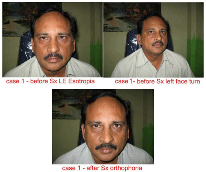
Figure 1:
Case 1 before and after surgery.
Case 1
Diagnosis: Left eye DRS with Esotropia
O/E: Left eye abduction limited, left face turn, FDT strongly positive for medial rectus, primary deviation 20 prism diopters, secondary deviation 40 prism diopters
Surgery: Left medial rectus recession 5mm
Result: Orthophoria in primary position
In many cases the unilateral medial rectus recession can eliminate the ET in primary position [4]. In two situations however, a recession of the contralateral medial rectus should also be done.
One example is DRS with ET for which the surgery is to include a large ipsilateral lateral rectus recession to treat severe retraction (see discussion on retraction).
A second example where a bilateral medial rectus recession should be considered is a subtype of DRS with ET over 20 PD in which there is limited adduction and very slow adduction saccadic velocities in the affected eye. In patients with DRS with limited abduction and an ET of 20 to 25 or more, a large medial rectus recession, up to 6 or 7mm has been recommended [2,4,5]. If adduction saccadic velocities are very slow and limitation of adduction is present, one should consider limiting the recession of the medial rectus of the affected eye to no more than 5 mm and adding a recession of the opposite medial rectus to fully correct the ET.
Duane Syndrome with XT (Table 1), A patient with XT requires a weakening of the ipsilateral lateral rectus in the surgical plan [2,4-6](Figure 2). If the retraction is severe and an abnormal head position is present due to the incomitant horizontal deviation, this procedure can also relieve these anomalies. If the XT is over 25 PD, consideration should be given to including a recession of the contralateral lateral rectus (Figure 3).
-
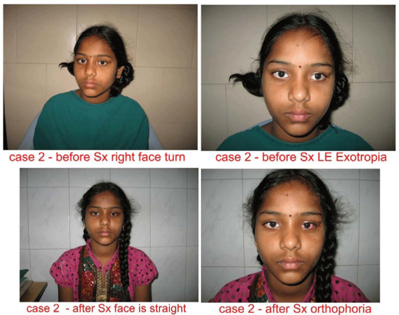
Figure 2:
Case 2 before and after surgery.
-
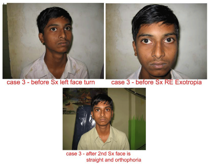
Figure 3:
Case 3 before and after surgery.
Case 2
Diagnosis: Left eye DRS with exotropia
O/E: Right face turn, exotropia of 20 prism diopters
Surgery: Left lateral rectus recession 8mm
Result: Face turn corrected and orthophoria
Case 3
Diagnosis: Right eye DRS with exotropia
O/E: left face turn150, primary deviation 25 prism diopters and secondary deviation 40 prism diopters exotropia
1st surgery: Right lateral rectus recession 9mm
Result: residual exotopia, primary deviation 15prism diopters, secondary deviation 25prism diopters exotropia and residual left face turn
2nd surgery: Left lateral rectus recession 9mm on adjustable suture
Result: Corrected compensatory head posture, Orthophoria in primary position
Forced duction testing must be done at the start of surgery and periodically during the operation as the various tissue planes are entered and recessed. If restrictions have not been sufficiently released to normalize forced ductions, then the amount of recession of a contractured muscle may have to be adjusted during surgery.
Abnormal Head Positions
Abnormal head postures are frequently seen in DRS [7,8]. A patient with an incomitant strabismus will adopt a head posture to obtain comfortable single binocular vision. If there is an imbalance of muscle forces in the primary position, the patient will select a field of gaze where the affected eye has a balance of forces [2,9,10].
In DRS with ET and limited abduction, a face turn toward the side of the affected eye is often noted (Figure 1). A medial rectus contracture is commonly found due to an imbalance of the forces of the medial rectus-lateral rectus agonist-antagonist muscle pair. Conversely, in DRS with XT and markedly limited adduction, it if the lateral rectus that often undergoes contracture then a face turn to the side opposite the affected eye may result (Figures 1 and 3).
The surgical plan must include releasing of horizontal restrictions, including muscle contractures.
Retraction of the globe
Electrophysiologic studies of DRS confirm that the lateral rectus innervation pattern is abnormal in almost all cases, leading to co-contraction with the medial rectus on attempted adduction [5,11].The resulting pseudoptosis that is observed clinically is rarely due to orbicularis or levator inhibition [3,6-8]. In this study, retraction was considered "severe" if there was at least a 50% narrowing of the center of the palpebral fissure on attempted adduction as compared to the primary position (Table 2).
-
Table 3:
Approach for Treatment of Upshoots and Downshoots.
LR - lateral rectus, SR = Superior Rectus, IR = Inferior Rectus
In order to reduce retraction in severe cases where the lateral rectus contributes a strong force either by significant co-contraction or by abnormal stiffness, the muscle must be recessed. In a case of DRS with XT, the recession of a tight, fibrotic muscle of 7 or 8 mm can significantly reduce the retraction and, at the same time, reduce the deviation [2,4,10]. In cases of DRS with XT in which the lateral rectus appears structurally normal at surgery or in cases of DRS with ET, retraction of a significant degree can be relieved with a larger recession of the muscle. It should be recessed 10 to 12 mm or more (Figure 5). If a non-fibrotic muscle is not recessed more than 7 or 8 mm in such cases, the abnormally co-contracting muscle can still "take up its slack" and a cosmetically poor enophthalmos on adduction can recur.
Upshoots and Downshoots
These abnormal movements are frequently seen in DRS and are generally associated with A, V or X patterns [4,6,7,11-13]. Several authors have addressed the etiology of these vertical movements both from mechanical and innervational considerations (Table 3).
Mechanical causes
The mechanical cause for upshoots and downshoots has been attributed to the side-slip of a tight lateral rectus as the adducted globe moves above or below the horizontal plane [10,13,14]. This has been termed a "bridle" effect [10,14]. The upshoots and downshoots caused by side-slip of the lateral rectus can be improved by several options. One approach is to recess the muscle. The dosage of recession is determined by the stiffness of the lateral rectus on forced ductions and whether the muscle is found to be fibrotic on examination at surgery. Recession a very stiff, fibrotic muscle 7 or 8 mm can significantly reduce the bridle effect and correct an XT of up to 20 to 25 PD in primary position. A non-fibrotic lateral rectus with mildly positive forced ductions should be recessed more, at least 10 to 12mm, to achieve the same goal. If the appropriate amount of surgery is performed, any pre-existing A, V, or X pattern should be significantly reduced or eliminated.
* Alternatives include Y-splitting or posterior fixation suture
Alternative surgical approaches include: posterior fixation (Faden) sutures on the lateral rectus, with or without similar sutures on the ipsilateral medial rectus, along with appropriate recessions of these muscles [13,14], and splitting of the lateral rectus tendon into a Y configuration (Figure 4) [15].
-
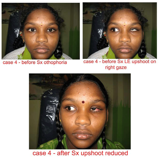
Figure 4:
Case 4 before and after surgery.
Case 4
Diagnosis: Left eye DRS with upshoot and globe retraction on right gaze
O/E: Orthophoria in primary position, palpebral fissure changes and upshoot of left eye on right gaze
Surgery: "Y" splitting of left lateral rectus
Result: Palpebral fissure changes and upshoot were reduced
Innervational Causes: Innervational anomalies may explain some cases of anomalous vertical movements. There is EMG evidence for co-contraction of superior rectus, with or without the inferior oblique, with the lateral rectus associated with upshoots, and for inferior rectus co-contraction with the lateral rectus in cases of downshoots [3,5,11,12]. Neuropathologic and neuroimaging studies have shown the lateral rectus to be innervated by a branch of the third cranial nerve in DRS. These results suggest that combinations of vertical rectus muscle-lateral rectus muscle co-innervation should be responsible for upshoots and downshoots in at least some cases.In these patients, preoperative magnetic resonance imaging should be done to study the relationship of extra ocular muscles with third nerve.The anomalous movements should respond to weakening the appropriate vertical muscle(s) along with appropriate horizontal muscle surgery (Figure 5).
-
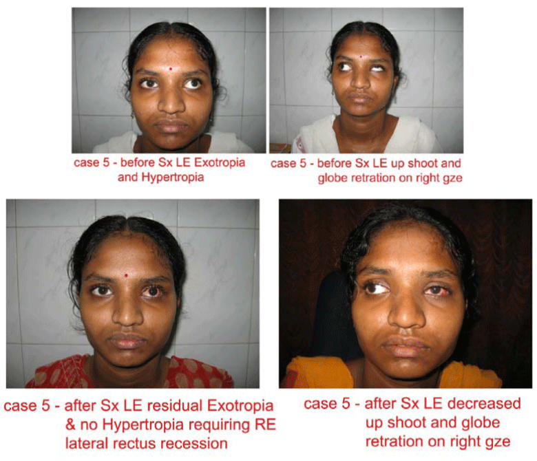
Figure 5:
Case 5 before and after surgery.
Case 5
Diagnosis: Left eye DRS with Exotropia and Hypertropia
O/E: Severe globe retraction and gradual upshoot o right gaze, 30 prism diopters exotropia and 20 prism diopters hypertropia
Surgery: left lateral rectus recession 15 mm and left superior rectus recession 7mm after adjustable suture
Result: Globe retraction, uphoot on right gaze are decreased, but there is residual exotropia of 15 prism diopters requiring right lateral rectus recession in second surgery.
Two clinical findings in a case of DRS with anomalous vertical movements may indicate that the patient falls into the subgroup where there is primarily an innervational cause. The first sign is the pattern of a gradual movement of elevation or depression. The vertical deviation steadily increases as the affected eye moves from abduction to primary position and into adduction. By contrast, in the mechanical form the eye usually remains in the horizontal plane or close to it, as the eye moves into adduction. And the flip-up or flip-down is typically abrupt and occurs as the eye rises just above or depresses just below the horizontal plane [10,14,15].
Case - 5 shows a pattern of gradual upshoot with a large vertical tropia in primary position, suggesting an innervational cause. It illustrates the fact that in carefully selected cases, the weakening of a vertical rectus muscle can significantly relieve an anomalous vertical movement, as well as retraction, in DRS without overcorrecting the vertical deviation in the primary position.
Bilateral duane syndrome
This entity represents up to 20% cases of DRS [1,4,7,10,16]. There are no large series describing consistent approaches to or results of treatment in this disorder, primarily because there are so many variations of presentation. Cases can be symmetric with mild or severe retraction, and the primary position deviations can range from small (15 to 20 PD) to large (60 to 70 PD) [4] (Figure 6). Cases can also be very asymmetric, and the two eyes can have two different forms of DRS (eg: one with limited abduction and the other with limited adduction) [4,7].
-
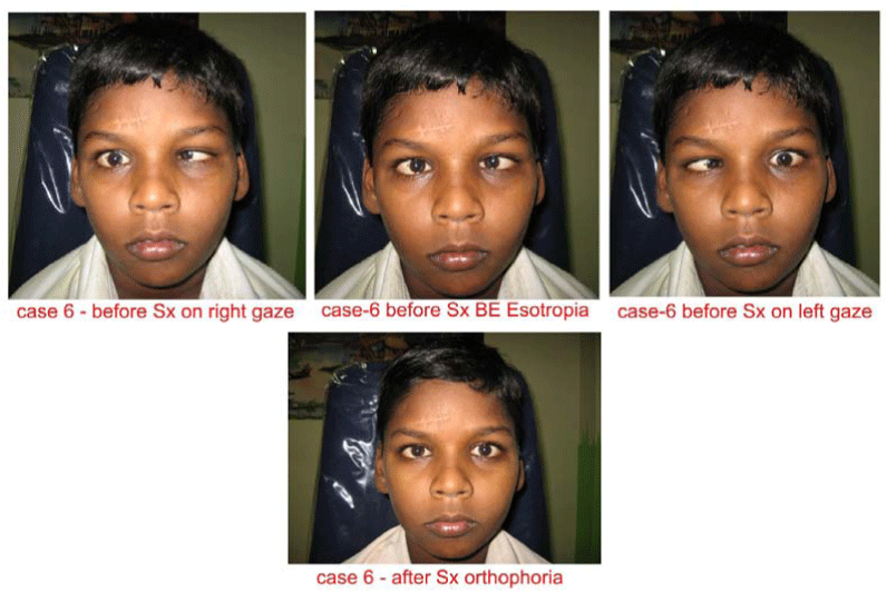
Figure 6:
Case 6 before and after surgery.
Case 6
Diagnosis: Bilateral DRS with esotropia
O/E: Limited abduction both eyes, FDT positive for both medial rectii, Deviation > 50 prism diopters with fixing right eye or left eye
Surgery: Bilateral medial rectus recession 6 mm each eye
Result: Orthophoria in primary position
Because there is such variability in presentation of bilateral DRS, each case must be approached on an individual basis. It is probably advisable to consider the correction of any face turn as the primary goal of surgery. As discussed earlier, this requires appropriate surgery on the preferred eye, as it "governs” the head position. The second consideration is the deviation in primary position. While the surgery on the preferred eye may simultaneously help correct the misalignment, in many cases one should be prepared to work on muscles in the other eye, as well as to totally eliminate the angle.
Discussion
There has been an emphasis in recent years on categorizing DRS (eg: Types I, II and III based upon the pattern of limitations of ductions in the affected eye [4-7]. Huber attempted to justify such a scheme bases upon EMG data [12]. However, clinically there is actually a spectrum of these patterns in DRS. Many cases have intermediate ranges of duction limitations causing them not to fall nicely into one of the "pure” categories [3,7].
Other authors have carefully avoided such dogmatic categorizations [1,6,8,17]. Instead, they pointed out the variability of findings and used other parameters to analyze their data. In planning surgery on a given case, it is best not to adhere to pure classifications, as this limits versatility in planning the optimal surgical procedure for that patient.
It is hoped that the approach listed here will be of assistance in selecting an appropriate surgical plan for a given case of DRS. Before surgery, co-existing significant refractive errors, anisometropia, and amblyopia must be treated. In addition, systemic anomalies associated with DRS [7,16] should be noted, and appropriate referral to medical consultants may be indicated in the full evaluation of patients.
Conclusion
Satisfactory cosmetic results can be achieved after operating Duane retraction syndrome in terms of horizontal squint, upshoot / downshoot, retraction of globe or abnormal head position.
Acknowledgement
Stephen P Kraft (Canada), David L Guyton (USA).
-
-
- Isenberg S, Urist MJ (1977) Clinical observation in 101 consecutive patients with Duane's retraction syndrome. Am J Ophthalmol 84:419-425.
- Feretis D, Papastratigakis B, Tsamparlakis J (1981) Planning surgery in Duane's Syndrome. Ophthalmologica183:148-153.
- Papst W, Esslen E (1964) Symptomatology and therapy in ocular motility disturbances. Am J Ophthalmal 58:275-291.
- Pressman SH, Scott WE (1986) Surgical treatment of Duane's syndrome. Ophthalmology 93:29-38.
- Metz HS, Scott AB, Scott WE (1975) Horizontal saccadic velocities in duane's syndrome. Am J Ophthalmol 80:901-906.
- Sato S (1960)Electromyographic study on retration syndrome. Jpn J Opthalmol 4:57-66.
- Breinin GM (1957)Eletromyography – A tool in ocular and neurologic diagnosis: II, Muscle palsies. Arch Ophthalmol 57:165-175.
- Spaeth EB (1953)Surgicalaspects of defective abduction. AMA Arch Ophthalmol 49:49-52.
- MacDonald AL, Crawford Js, Smith DR (1974) Duane's retraction syndrome: an evaluation of the sensory status. Can J Ophthalmol 9:458-462.
- Jampolsky A (1978)Surgical leashes and reverse leashesin strabismus surgical management, in symposium on Strabismus: Transactions of the New Orleans Academy of ophthalmology. St Louis CV Mosby 244-268.
- Scott AB, Wong G, Jampolsky A (1970) Pathogenesis in Duane's syndrome(abstract). Invest Ophthalmol Vis Sci9:983.
- Huber A (1974) Electrophysiology of retraction syndromes. Br J Ophthalmol58: 293-300.
- Scott AB (1978)Upshoots and downshoots, in Souza-Dias C (ed): V. Congress of C.L.A.D.E. (Conselho Latino-Americano de Estrabismo), October 16-17, 1976, Guaruja-Brasil. Sao Paulo, Oficinas das Edicoes Loyola 60-65.
- Von Noorden GK, Murray E (1986) Up and down shoot in Duane's retraction syndrome. J Pediatr Ophthalmol Strabismus 23:212-215.
- Roger s GL, Bremer DL (1984)Surgical Treatment of the upshot and downshoot in Duane's Retraction syndrome. Ophthalmology91:1380-1383.
- O'Malley ER, Helveston EM, Ellis FD (1982) Duane's retraction syndrome – Plus. J Pediatr Ophthamol Strabismus19:161-165.
- Blodi FC, Van allen MW, Yarbrough JC (1964) Duane's syndrome; A brain stem leision. Arch Ophthalmol 72:171-177.
-
-


Table 1:
Approach to Treatment of Primary Position Deviation.
PD-prism diopters, ET-esotropia, XT-exotropia, MR-medial rectus, LR-lateral rectus, OU-both eyes
Table 1: Approach to Treatment of Primary Position Deviation.
PD-prism diopters, ET-esotropia, XT-exotropia, MR-medial rectus, LR-lateral rectus, OU-both eyes